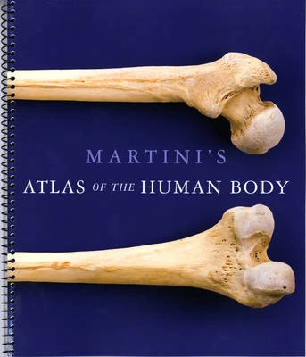
Martini's Atlas of the Human Body (ME Component); Frederic H Martini; 2011
2 säljare

Martini's Atlas of the Human Body (ME Component) Upplaga 9

av Frederic H Martini
The Atlas supplements the textbook with an abundant collection of anatomy photographs (194), radiology scans (53), and embryology summaries (21). The anatomy photographs, taken by Ralph Hutchings, the renowned biomedical photographer, extend the anatomy photographs that appear in the book. The radiology scans help students visualize deep anatomical structures and encourage them to become familiar with the types of images seen in a clinical setting. The embryology summaries depict the developmental origins of major tissues, organs, and body systems. This is the ME Component item that is automatically packaged with the student text.
Upplaga: 9e upplagan
Utgiven: 2011
ISBN: 9780321724564
Förlag: Pearson
Format: Bok
Språk: Engelska
Sidor: 160 st
The Atlas supplements the textbook with an abundant collection of anatomy photographs (194), radiology scans (53), and embryology summaries (21). The anatomy photographs, taken by Ralph Hutchings, the renowned biomedical photographer, extend the anatomy photographs that appear in the book. The radiology scans help students visualize deep anatomical structures and encourage them to become familiar with the types of images seen in a clinical setting. The embryology summaries depict the developmental origins of major tissues, organs, and body systems. This is the ME Component item that is automatically packaged with the student text.
Begagnad bok
145 kr
Fri frakt & skickas inom 1-3 vardagar
Köpskydd med Studentapan
Varje köp täcks av Studentapans köpskydd som säkerställer att boken kommer fram, att du får rätt bok och att skicket stämmer överens med beskrivning.
145 kr
Fri frakt & skickas inom 1-3 vardagar