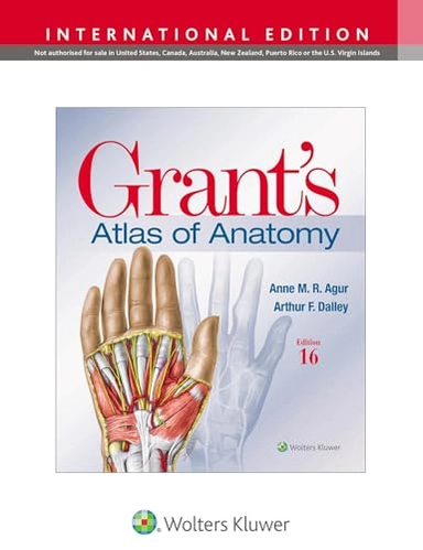
Grant's Atlas of Anatomy; Anne M R Agur, Arthur F Dalley Ii; 2024
endast ny

Grant's Atlas of Anatomy Upplaga 16

av Anne M R Agur, Arthur F Dalley Ii
Illustrations drawn from real specimens, presented in surface-to-deep dissection sequence, set Grant’s Atlas of Anatomy apart as the most accurate illustrated reference available for learning human anatomy and referencing in dissection lab. A recent edition featured re-colorization of the original Grant’s Atlas images from high-resolution scans, also adding a new level of organ luminosity and tissue transparency. The dissection illustrations are supported by descriptive text legends with clinical insights, summary tables, orientation and schematic drawings, and medical imaging. Renowned, high-resolution, dynamically colored illustrations organized in dissection sequence enable the formation of 3D constructs for each body region and provide detailed, realistic reference during dissection. Tables detail muscles, vessels, and other anatomic information in an easy-to-use format ideal for review and study. Enhanced medical imaging includes more than 100 clinically significant MRIs, CT images, ultrasound scans, and corresponding orientation drawings to help students confidently apply the laboratory experience to clinical rotations. Color schematic illustrations reinforce the relationships of structures and anatomical concepts in vibrant detail.
Upplaga: 16e upplagan
Utgiven: 2024
ISBN: 9781975193447
Förlag: Wolters Kluwer Health
Format: Häftad
Språk: Engelska
Sidor: 896 st
Illustrations drawn from real specimens, presented in surface-to-deep dissection sequence, set Grant’s Atlas of Anatomy apart as the most accurate illustrated reference available for learning human anatomy and referencing in dissection lab. A recent edition featured re-colorization of the original Grant’s Atlas images from high-resolution scans, also adding a new level of organ luminosity and tissue transparency. The dissection illustrations are supported by descriptive text legends with clinical insights, summary tables, orientation and schematic drawings, and medical imaging. Renowned, high-resolution, dynamically colored illustrations organized in dissection sequence enable the formation of 3D constructs for each body region and provide detailed, realistic reference during dissection. Tables detail muscles, vessels, and other anatomic information in an easy-to-use format ideal for review and study. Enhanced medical imaging includes more than 100 clinically significant MRIs, CT images, ultrasound scans, and corresponding orientation drawings to help students confidently apply the laboratory experience to clinical rotations. Color schematic illustrations reinforce the relationships of structures and anatomical concepts in vibrant detail.
Ny bok
876 kr922 kr
5% studentrabatt med Studentapan
Begagnad bok (0 st)
Ny bok
876 kr922 kr
5% studentrabatt med Studentapan
Begagnad bok (0 st)